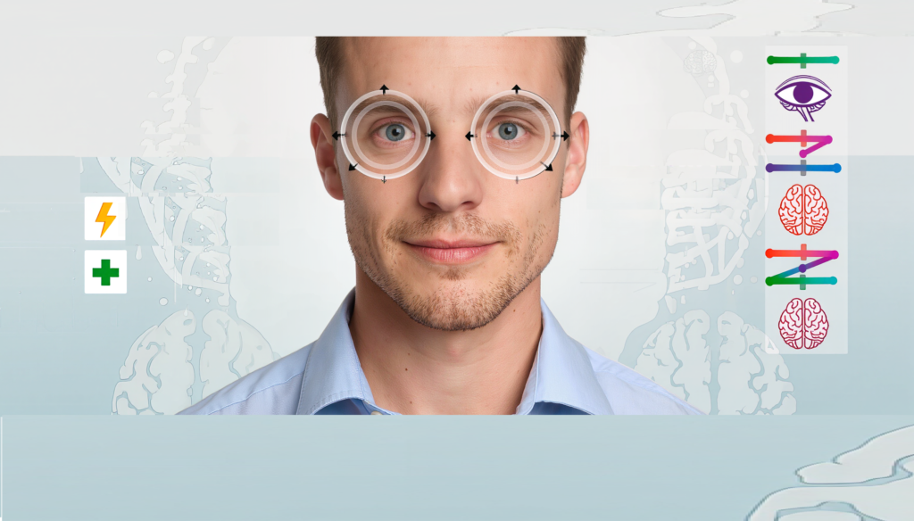












Your pupils are more than just the black centers of your eyes. They are tiny windows into your brain and nervous system. The way they constrict and dilate in response to light can reveal a surprising amount about your neurological health.
Doctors have long used pupil size and reaction as part of a neurological exam. But recent research shows that even subtle changes in pupil behavior—like uneven dilation or delayed response to light—can be early warning signs of serious conditions, from brain injury and stroke to autonomic nervous system dysfunction.
In this article we’ll explore how pupil dilation works, what abnormal patterns can signal, and why this simple sign is becoming an increasingly important tool in modern neurology.
How Your Pupils Work: The Autonomic Connection
Your pupils are controlled by the autonomic nervous system (ANS), which regulates involuntary bodily functions like heart rate, digestion, and yes, pupil size. Two branches of the ANS work in opposition:
- Sympathetic nervous system: Causes pupil dilation (mydriasis) in response to stress, darkness, or arousal.
- Parasympathetic nervous system: Causes pupil constriction (miosis) in response to light or near focus.
This balance is called the pupillary light reflex (PLR). When light hits your retina, signals travel to the brainstem, which then tells the pupils to constrict. When light decreases, the pupils dilate.
The PLR is a sensitive, non-invasive measure of autonomic function. Any disruption in this reflex can point to damage in the brainstem, cranial nerves, or autonomic pathways.
What Is Anisocoria And Why Does It Matter?
Anisocoria is the medical term for unequal pupil size. Normally, both pupils constrict and dilate equally in response to light. When they don’t, it can signal a problem.
Anisocoria can be:
- Physiological: A normal, harmless variation in some people (about 20% of the population).
- Pathological: A sign of neurological or eye disease.
The key is context. A slight difference in pupil size that is stable over time and has no other symptoms is often benign. But a new or changing difference—especially with other symptoms—can be a red flag.
How Doctors Interpret Pupil Asymmetry
When a doctor notices unequal pupils, they look at two things:
- Which pupil is abnormal?
- If the difference is greater in bright light, the larger pupil is abnormal (it is failing to constrict).
- If the difference is greater in dim light, the smaller pupil is abnormal (it is failing to dilate).
- How do the pupils react to light?
- If both pupils react normally to light, the smaller pupil is likely the problem (sympathetic pathway issue, like Horner syndrome).
- If one pupil does not react well to light, the larger pupil is likely the problem (parasympathetic pathway issue, like third nerve palsy).
This simple logic helps doctors narrow down the cause quickly.
Horner Syndrome: When The Small Pupil Is The Problem
Horner syndrome is a condition where one pupil is smaller than the other (miosis), often with:
- Mild drooping of the eyelid (ptosis).
- Decreased sweating on the same side of the face (anhidrosis).
- Delayed dilation in the dark.
It happens when the sympathetic nerves that control pupil dilation are damaged. Causes can include:
- Stroke or tumor affecting the brainstem or neck.
- Carotid artery dissection (a tear in the artery wall).
- Lung tumor (especially at the top of the lung, called a Pancoast tumor).
- Trauma or surgery affecting the neck or chest.
A key sign is dilation lag: the affected pupil takes longer to dilate in the dark compared to the normal pupil.
Horner syndrome can be a warning sign of serious underlying conditions, so it always warrants further investigation.
Third Nerve Palsy: When The Large Pupil Is The Problem
Third cranial nerve (oculomotor nerve) palsy affects the nerve that controls most eye movement and pupil constriction. When this nerve is damaged, you may see:
- A large, dilated pupil that does not react well to light.
- Drooping eyelid (ptosis).
- Eye turning outward and downward (because other muscles take over).
- Double vision (diplopia).
This is a medical emergency because it can be caused by:
- Aneurysm: Especially of the posterior communicating artery, which can compress the nerve.
- Brain tumor or hemorrhage.
- Increased intracranial pressure.
If you or someone else suddenly develops a dilated, non-reactive pupil with a drooping eyelid or double vision, seek emergency care immediately.
Pupillary Light Reflex And Brain Injury
In emergency settings, the pupillary light reflex is a critical tool for assessing brain function, especially after trauma.
How it works in trauma assessment
- Normal PLR: Suggests intact brainstem function and no significant pressure on the brain.
- Sluggish or absent PLR: Can indicate increased intracranial pressure, brainstem compression, or severe brain injury.
Dynamic pupillometry (measuring pupil response with a handheld device) is now used in ICUs and emergency departments to:
- Detect early signs of brain swelling.
- Monitor response to treatment.
- Predict outcomes in traumatic brain injury.
One study found that changes in pupil reactivity can predict worsening brain injury before other symptoms appear, making it a valuable early warning tool.
Pupil Abnormalities And Autonomic Dysfunction
Beyond acute emergencies, pupil behavior can reveal chronic autonomic nervous system problems.
Parkinson’s disease
Research shows that people with Parkinson’s disease often have:
- Reduced pupil constriction in response to light.
- Delayed pupil response times.
- Other signs of autonomic dysfunction.
This is because Parkinson’s affects the autonomic nervous system, which also controls pupil size.
Diabetes and other neuropathies
Diabetes can damage the nerves that control pupil function, leading to:
- Abnormal pupil reactions.
- Reduced sensitivity to light changes.
Other conditions
Pupil abnormalities have also been linked to:
- Multiple sclerosis.
- Alzheimer’s disease.
- Certain toxic exposures.
- Medication side effects.
How Doctors Test Pupil Function
A basic pupil exam is quick and painless. Here is what a doctor typically does:
- Measure pupil size in both light and dark. This helps determine which pupil is abnormal.
- Test the light reflex. Shine a light in each eye and watch for constriction.
- Check for afferent pupillary defect. This tests if the problem is in the optic nerve or retina.
- Assess eye movements and eyelid position. This helps identify cranial nerve involvement.
- Consider pharmacological testing. In some cases, special eye drops (like cocaine or apraclonidine) are used to confirm Horner syndrome.
For more detailed assessment, dynamic pupillometry can be used. This device measures:
- Pupil size over time.
- Speed of constriction and dilation.
- Latency (delay) in response.
This gives a much more precise picture of autonomic function than a simple flashlight test.
When To Seek Medical Attention
Not every case of unequal pupils is an emergency. But some situations require immediate care:
Seek emergency care if you have:
- A suddenly dilated pupil with:
- Drooping eyelid.
- Double vision.
- Eye pain.
- Headache.
- A new difference in pupil size after:
- Head injury.
- Loss of consciousness.
- Severe headache.
- Pupil changes with:
- Weakness or numbness on one side of the body.
- Confusion or altered mental state.
- Difficulty speaking.
See a doctor soon (but not necessarily emergency) if:
- You notice a persistent difference in pupil size.
- You have other symptoms like drooping eyelid or decreased sweating.
- You have a history of migraines or other neurological conditions.
The Future Of Pupil-Based Diagnostics
Pupillometry is becoming an increasingly important tool in medicine. Researchers are exploring its use in:
- Early detection of Alzheimer’s and other dementias.
- Monitoring of autonomic function in diabetes.
- Assessment of pain and stress levels.
- Detection of concussion and mild traumatic brain injury.
Because it is non-invasive, quick, and objective, pupillometry may become a routine part of neurological exams in the future.
Bottom line
Your pupils are more than just a cosmetic feature. They are a real-time readout of your brain and autonomic nervous system.
Key takeaways:
- Pupil size and reaction are controlled by the autonomic nervous system.
- Anisocoria (unequal pupils) can be normal or a sign of disease.
- Horner syndrome (small pupil) can signal problems in the sympathetic pathway.
- Third nerve palsy (large, non-reactive pupil) can be a sign of a brain aneurysm or other emergency.
- Pupillary light reflex is a valuable tool in assessing brain injury and autonomic function.
If you notice a new or changing difference in your pupil size, especially with other symptoms, don’t ignore it. It could be your body’s way of telling you something important.
Sources: